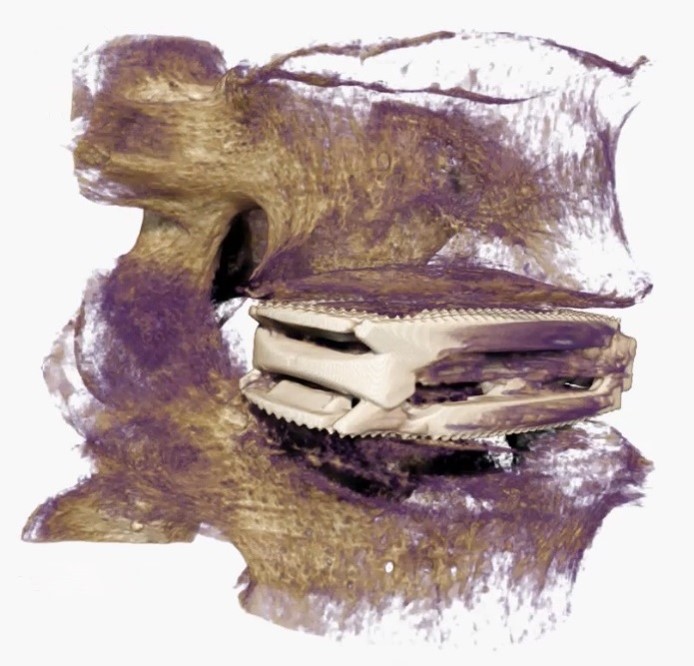
While conventional CT imaging produces images with a slice thickness of roughly 1mm, MicroCT is capable of resolving detail in the micrometre (μm) range. This vast improvement in resolution provides a level of detail never before seen with computed tomography. The image above was taken from a MicroCT scan of an interbody fusion cage being studied for its biomechanical properties in the lumbar spine. Each micrometre slice can be analyzed individually or viewed in its entirety. Fusion was subsequently quantified through MicroCT, histology, and manual palpation.