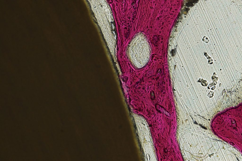
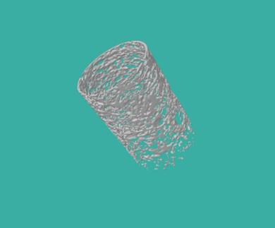
The image above was acquired from a 3D MicroCT volume, and represents a bioactive dowel used in an in-vivo investigational study of various biomaterials (ovine model). The substance being quantified is new bone around the dowel’s surface. Micro CT volumetric data is followed by direct histologic quantification of new bone growth area.